Por el Dr. Robert Lowe
El siguiente caso demuestra cómo colocar una restauración mínimamente invasiva utilizando el modo de detección de caries de una cámara intraoral y ACTIVA BioACTIVE-RESTORATIVE.
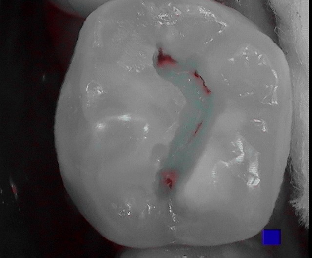
La figura 1 es una vista oclusal del diente número 29 tomada con el modo de detección de caries de una cámara intraoral (SoproCare: Acteon USA) que muestra una caries activa en la fisura central del diente. Es importante señalar que esta lesión aún no puede «pegarse» con un explorador debido a la profundidad de esta estrecha fisura y a la incapacidad de la punta del explorador para alcanzar la lesión debido a su mayor tamaño. Se utiliza una fresa de fisurotomía (micro NTF 6066: SS White) para erradicar cuidadosamente sólo la zona que presentaba fluorescencia positiva (aspecto clínico de mancha).
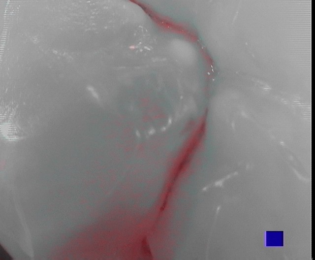
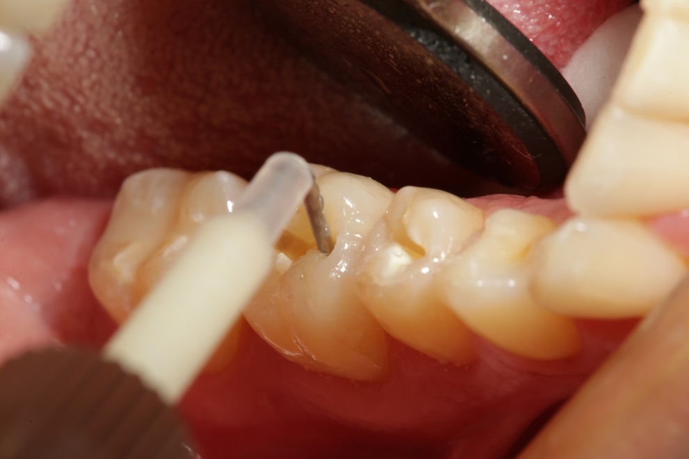
Mientras se realiza la preparación, se puede utilizar la cámara intraoral en modo de detección de caries (Cario) para comprobar la eliminación completa de la caries activa (Figura 2).
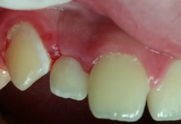
Como ACTIVA favorece el proceso natural de remineralización con liberación de calcio y fosfato, las zonas «blancas» del esmalte descalcificado pueden conservarse en lugar de eliminarse durante la preparación. Tras utilizar un protocolo de grabado total, colocación de adhesivo y fotopolimerización, ACTIVA BIOACTIVE-RESTORATIVE se coloca en la micropreparación utilizando su punta automix con una cánula flexible (Figura 3).
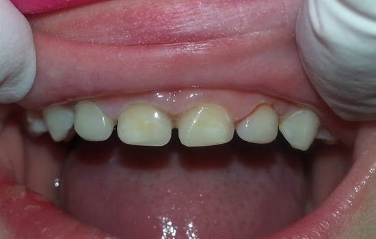
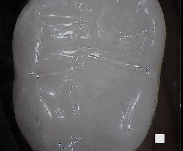
La figura 4 muestra una vista de la restauración terminada tomada con la cámara intraoral. Observa que es difícil, si no imposible, detectar la restauración incluso con un aumento extremo. Los márgenes de la restauración son imperceptibles. El diente parece intacto, como un esmalte virgen.
Referencias
- Ferracane JL, Pfeifer CS, Bertassoni LE, «Biomateriales para la salud bucodental», Dental Clinics of North America, vol.61, no.4, octubre de 2017, pp. 651-872.
- Chao W, et al. Desviación a la rotura de materiales de restauración. J Dent Res 94 (Spec Iss A) 2375, 2015 (iadr.org).
- Slowikowski L, et al. Liberación y recarga de iones fluoruro a lo largo del tiempo en tres materiales de restauración. Ponencia presentada en: AADR Annual Meeting & Exhibition 2014; 19 de marzo de 2014; Boston, MA.
- Garcia-Gadoy F, Morrow BR, Pameijer CH, «Flexural Strength and Fatique of Activa RMGICs», Facultad de Odontología UTHSC, Memphis y Facultad de Odontología UConn, Farmington, CT, Presentación de libro blanco en IADR, 2014.
- Maj J, Merritt J, Ferracane J, «Adhesion of S. Mutans Biofilms on Potentially Antimicrobial Dental Composites», J dent Res 96 (Special Issue A); 2560, 2017.
- Comba A, Breschi L, et.al., J Dent Res 97 (Special Issue A) 0273, 2018.
- Girn VS, et.al., J Res Dent 93 (Edición especial A): 1163, 2014.

Acerca de Robert A. Lowe, DDS
Robert A. Lowe, DDS, obtuvo el título de Doctor en Cirugía Dental en la Facultad de Odontología de la Universidad de Loyola. Tras completar su residencia, el Dr. Lowe pasó a la práctica privada y empezó a dedicarse a otra pasión: la enseñanza clínica. Mientras dirigía su propia consulta, el Dr. Lowe ejerció como Profesor Clínico de Odontología Restauradora en la Facultad de Odontología de la Universidad de Loyola hasta su clausura en 1993. En 2000, se trasladó a Charlotte, Carolina del Norte.